The preliminary investigation
In order to ensure maximum safety and precision during the operation, a comprehensive and precise preliminary examination is of great importance. As part of this preliminary examination, numerous measurements and examinations are carried out that are necessary for the precise fitting of the artificial lens.
The pupil will also be dilated with medication. Therefore, please note that you must not drive for 3 hours after this examination. Ideally, you should be collected by someone you trust. A medical report from your GP is very helpful if you have to take medication regularly or if other important treatments are required on a regular basis.
The day of the operation
On the day of the procedure, please have breakfast as normal and take your medication as usual. However, please refrain from wearing make-up, face creams, perfume or eau de toilette.
The local anesthetic
The eye is usually anesthetized with eye drops during the operation. This minimizes the stress of the operation and the effect wears off quickly after the procedure. A low-dose sedative can also be used in addition. In rare cases, an injection next to the eye or a general anaesthetic can also be administered if absolute immobilization of the eye is required.
Preparation
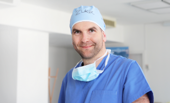
After dilating the pupil with drops or inserting a tablet into the palpebral fissure, the patient is led into the operating theater wearing special surgical clothing (gown, cap, etc.).
The patient is placed on the operating table, then the operating area is neatly covered with sterile drapes and foil. A sufficient oxygen supply is of course guaranteed, but this is monitored by means of a pulse oximeter during the entire stay in the operating theater.
An eyelid holder is then inserted to keep the eye open during the procedure. To prevent the surface of the eye from drying out, the eye is permanently moistened with a special liquid.
The OP
The operation is performed under a high-resolution microscope. A tiny incision is made in the eye with a steady hand. The lens capsule, which encloses the cortex and the nucleus like a shell, is opened through this incision. The cloudy lens can now be broken up and suctioned out using ultrasound waves. What remains is the empty lens capsule – the so-called capsular bag.
The tightly rolled-up lens is now inserted into the eye through the small incision, where it can slowly unfold in the capsular bag.
Finally, the wounds are sealed and an antibiotic is injected into the anterior chamber to prevent infection.
The eye is then treated with an eye cup.