Elevated eye pressure: symptoms, treatment, causes and risks of high intraocular pressure
A little pressure never hurts – they say. Does that also apply to the eye? Not quite. Increased intraocular pressure can go unnoticed, but can have serious long-term consequences for your eyesight – including glaucoma. In this article, you will find out how eye pressure develops, what symptoms and risks are associated with it – and why it plays a crucial role if you are considering laser eye treatment or lens surgery. Clear, understandable and with an eye for the essentials.
What does increased eye pressure mean – and when does it become a problem?
Increased eye pressure occurs when the aqueous humor in the eye is no longer in balance – in other words, either too much is produced or too little is removed. This clear fluid system circulates inside the eye, more precisely between the lens, iris and the so-called chamber angle, where the outflow takes place. If this is disturbed, the pressure in the eye rises.
How high should the intraocular pressure normally be?
Normally, intraocular pressure is between 10 and 21 mmHg. Values above this are considered elevated – not always dangerous, but potentially risky. This is because high eye pressure can lead to damage to the optic nerve in the long term. Particularly problematic: the increased pressure usually causes no pain or symptoms – it often remains undetected for a long time.
It becomes critical when the pressure increases permanently, because:
- It can change the round shape of the eye.
- It strains the sensitive inner layers of the eye.
- It can lead to damage to the optic nerve – often irreversible.
In this context, ophthalmologists often speak of a risk factor for glaucoma – a serious eye disease that can permanently impair vision. Therefore, anyone who suspects increased intraocular pressure should definitely have an eye examination.
Causes of increased intraocular pressure
There are many possible causes of increased eye pressure – from harmless to absolutely worth investigating:
Imbalance in the outflow of aqueous humor
The most common cause of increased eye pressure is an imbalance between the production and drainage of aqueous humor. The aqueous humor is produced by the ciliary body behind the iris and circulates through the eye. It supplies the cornea and lens with nutrients before it drains through the angle of the eye.
If this outflow is disturbed – for example due to a narrowing of the chamber angle, adhesions or structural changes – the aqueous humor can no longer be removed to a sufficient extent. As a result, the pressure in the eye increases.
Typical causes of this impaired drainage are
- Predisposition: Especially in people with flat anterior chambers of the eye.
- Inflammations or injuries that block the chamber angle.
- Age-related changes in the eye tissue.
- Vascular changes or circulatory disorders
In rare cases, a certain enzyme that is responsible for the breakdown of metabolic products in the aqueous humor is even missing. This can also lead to an increase in pressure in the long term. If left untreated, there is a risk of gradual damage to the optic nerve, which can lead to impaired vision.
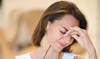
Symptoms and warning signs of high eye pressure
Elevated eye pressure often remains symptom-free for a long time – and this is precisely what makes it so insidious. In contrast to many other diseases, high intraocular pressure does not cause acute pain, redness or clearly noticeable changes. The first warning signs often only appear when damage to the optic nerve has already occurred.
Typical signs that may indicate high eye pressure:
- Blurred vision or circles of light around light sources (especially at night)
- Feeling of pressure in the eye (rare)
- Headaches, especially in the forehead area
- Visual deterioration without a recognizable cause
- Later: loss of visual field, which usually begins unnoticed
In particularly severe cases – such as an acute attack of glaucoma – severe eye pain, nausea, vomiting and a dilated, fixed pupil occur very suddenly. This is an ophthalmological emergency that must be treated immediately to prevent permanent damage to the optic nerve.
Diagnosis and measurement of eye pressure (tonometry)
The diagnosis of increased eye pressure begins with a precise measurement of the intraocular pressure – also known as tonometry. This examination is one of the most important routine ophthalmological checks and is completely painless.
There are various methods for measuring eye pressure, which are used differently depending on the practice:
- Applanation tonometry: The gold standard – the cornea is slightly flattened to determine the pressure in the eye.
- Non-contact tonometry (air blast measurement): Particularly popular for screening examinations as it works quickly and without contact.
- Rebound tonometry: Used primarily for children or sensitive patients.
In addition, many ophthalmologists also use supplementary diagnostics, e.g:
- OCT (optical coherence tomography) to visualize the optic nerve
- Visual field measurement (perimetry) to detect initial loss of function
- Ventricular angle analysis with special lenses or imaging procedures
This combination of tonometry, optic nerve monitoring and functional tests allows early signs of glaucoma to be detected – even before there is any measurable impairment of vision. If necessary, a good ophthalmologist will also regularly check the outflow of aqueous humor in order to better assess the causes and risks of a possible increase in pressure.
Treatment options for increased intraocular pressure:
If increased intraocular pressure is detected, the treatment depends on how high the pressure is, whether there is already damage to the optic nerve and the underlying cause. The aim of any treatment is to lower the eye pressure in order to protect vision in the long term and prevent or slow down the development of glaucoma.
1. drug treatment
The first step is usually the administration of eye drops, which:
- reduce the production of aqueous humor,
- improve the outflow via the chamber angle,
- or do both at the same time.
The common active ingredients include:
- Prostaglandin analogs (improve the outflow),
- Beta-blockers (reduce aqueous humor production),
- Carbonic anhydrase inhibitors (reduce production in the ciliary body),
- Alpha agonists (double effect).
2. laser treatments
If eye drops do not work sufficiently or are not tolerated, laser treatment may be useful:
- Laser trabeculoplasty: improves the outflow of aqueous humor.
- Iridotomy: for narrow chamber angles to improve pressure equalization.
3. surgical interventions
In advanced cases or if the increase in pressure is resistant to treatment, surgery may be considered:
- Trabeculectomy: creates a new drainage path for the aqueous humor.
- Microimplants (e.g. iStent): are inserted into the chamber angle to permanently improve the outflow.
4 Supplementary measures
A healthy lifestyle also plays a role:
- Blood pressure control
- Regular exercise
- Abstaining from smoking
- Avoid pressure on the eye, e.g. by pressing hard

Effects of high eye pressure on laser eye surgery / lens surgery
Increased eye pressure is an important aspect that must be taken into account when planning laser eye treatment or lens surgery. This is because intraocular pressure can affect suitability for certain procedures as well as safety and healing after the operation.
Why the eye pressure is carefully examined before the procedure:
Before a laser treatment (e.g. Femto-LASIK, Trans-PRK, SmartSight) or lens replacement is planned, a comprehensive diagnosis is always carried out, including measurement of the eye pressure. Increased pressure in the eye can indicate existing or incipient glaucoma – a condition that must be treated with particular caution.
Possible effects:
- Increased risk of complications: If the pressure is too high, delayed wound healing, pressure fluctuations or even deterioration of the optic nerve may occur after the operation.
- Limitation of suitability: Not all laser or lens procedures are recommended for increased intraocular pressure – depending on the condition of the chamber angle, the optic nerve and the inner layers of the eye.
- Long-term monitoring necessary: Even after the operation, the pressure must be checked regularly to rule out possible consequential damage.
What this means for patients:
- Ophthalmologic clarification in advance is mandatory – at EyeLaser Vienna & Linz, comprehensive diagnostics with tonometry and optic nerve analysis are therefore carried out before every refractive procedure.
- Individual consultation: If the eye pressure is elevated, we will check together whether surgery is still possible – possibly with an adapted technique or after prior medication.
- No automatic diagnosis of exclusion: An elevated intraocular pressure does not mean that surgery is not possible – but the decision must be based on sound diagnostics and experience.
So anyone considering laser eye treatment or lens implantation should definitely have their eye pressure checked at an early stage. This provides certainty – and creates the basis for individual and low-risk treatment.