The anatomy of the eye
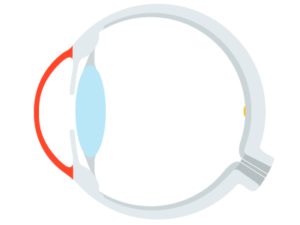
LThe eye is one of the most important human sensory organs and a complex optical system. The eye enables us to perceive the world in all its diversity, experience colors and recognize spatial structures. Vision works in a similar way to a highly sensitive camera: light passes through the camera lens and is focused by individual “components” of the eye – cornea, anterior chamber, pupil, lens and vitreous body – and imaged on the retina: the back wall of the eye.
In an eye with normal vision, the light rays meet exactly at a focal point on the retina, usually at the point of sharpest vision.
An image of our surroundings is only formed when the rays of light reach our eyes with the correct refraction. In people with normal vision, the total refractive power of the eye is balanced, which is why they do not need visual aids such as glasses or contact lenses. The total refractive power of the eye is measured in diopters (dpt).
A visual defect or visual impairment such as short-sightedness, long-sightedness or astigmatism occurs when the rays of light can no longer be optimally refracted, which means that the rays can no longer focus at one point. A distinction is made between short-sightedness, long-sightedness and astigmatism as well as presbyopia.
The cornea
The outer skin of the eye surrounds the entire eyeball as protection and consists of the sclera and cornea. The sclera takes up around 80 percent of the outer eye wall; we see the sclera as the white part of the eye.
The cornea, on the other hand, is transparent and forms the front part of the outer skin of the eye. The pupil can be seen through the cornea. This part of the eye is responsible for two thirds of the refractive power of the eye and is therefore very important for vision. The cornea is curved so that the light rays can be refracted and focused in a central point. If you suffer from defective vision, the cornea, which is not optimally curved, is often to blame.
iris (iris)
The iris forms the eye’s diaphragm. The iris surrounds the lens and is responsible for how much light passes through the pupil. Depending on how bright or dark it is, the iris narrows or widens. This work of the iris allows the eye to adapt to the respective light situation and regulate the depth of field.
Lens and ciliary body
The lens is located behind the pupil, between the vitreous body and the iris. The lens is responsible for the optimal focusing of light and for one third of the refractive power of the eye. The ciliary muscle, which is located in the ciliary body, has the task of adjusting the shape of the lens. Depending on the distance at which you want to see, the ciliary muscle adjusts the lens for the necessary sharpness. The ciliary muscle can stretch the lens flat, which reduces the refractive power of the light. If you want to see close up, the refractive power must be increased and the ciliary muscle shapes the lens back to its original curved shape.
The vitreous body
The vitreous body fills the inside of the eye between the lens and the retina and consists of a gel-like substance surrounded by a thin membrane.
The inner eye membrane (retina)
The inner skin of the eye consists of the pigment epithelium and the retina. The nerve cells with photoreceptors located in the pigment epithelium are responsible for color vision, among other things. Once the light has passed through the cornea, lens and vitreous body, it finally reaches the retina. Here, the light rays and all their information are converted into nerve impulses that are transmitted to the brain. It is only in the brain that the images received by the eye are converted into information. Like a computer, the nerve cells in the retina improve image contrast and color intensity and ensure that moving images are displayed more clearly.
Site of sharpest vision: the yellow spot (macula)
The yellow spot is located in the center of the retina. Due to the very high cell density of the photoreceptors, this is the point of sharpest vision. When we look at an object, our eyes automatically rotate so that the macula is always at the center of the light focus.
The optic nerve
The optic nerve transmits information from the retina to the brain. Like the retina, the optic nerve is also part of the brain.